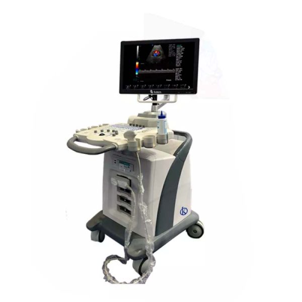
Using an ultrasound scanner in the medical line is crucial for obtaining accurate and swift diagnoses.
This article offers a detailed guide to making the most of this technology and focusing on the latest trends in medical technology and recent advances in laboratory research.
If you’re seeking a blend of innovation and quality, you’ve come to the right place. At https://kalstein.it/category-product/medical-line/ultrasound-scanner/ we offer you the luxury to explore our exclusive catalog of laboratory equipment. We manufacture each piece of equipment with a level of excellence. Our intuitive and agile online shopping channels are designed for your convenience, ensuring the friendliest prices. Don’t hesitate any longer, we bring science to life, it’s time to become part of our community. https://kalstein.it/
Initial Preparation: Setting Up the Ultrasound Scanner
Before starting any procedure, it’s essential to properly set up the ultrasound scanner. First, ensure that the equipment is securely connected to a stable power source and that all cables are firmly attached. The initial calibration of the device is crucial; follow the manufacturer’s instructions to ensure all settings are correctly adjusted.
Additionally, it’s important to clean and disinfect the ultrasound probes before each use. Use cleaning products recommended by the manufacturer to avoid damaging the equipment. Proper setup and equipment hygiene not only extend the scanner’s lifespan but also ensure the accuracy of the study results.
Choosing the Appropriate Probe for Each Procedure
Selecting the correct probe is crucial for obtaining high-quality images. There are different types of probes, each designed for specific applications. For example, linear probes are ideal for superficial exams such as thyroid or breast studies, while convex probes are better suited for abdominal and obstetric studies due to their greater penetration depth.
For vascular procedures, a high-frequency probe can provide more detailed images of blood vessels. Always consult the user manual and best practice guidelines to ensure you’re using the appropriate probe for the study in question. The correct probe choice maximizes diagnostic accuracy and enhances the quality of the medical service provided.
Scanning Techniques: Image Optimization
To achieve the best results with an ultrasound scanner, it’s essential to master scanning techniques. Start by applying a generous amount of ultrasound gel to the patient’s skin; this helps eliminate air between the probe and the skin, improving sound wave transmission. Place the probe on the patient’s skin and adjust the pressure as necessary to obtain clear images.
Adjust the scanner’s parameters, such as gain and depth, to optimize image quality. Gain controls the image brightness, while depth adjusts the distance at which sound waves are focused. Experiment with these settings during the procedure to achieve the best possible visualization. Proper scanning technique is essential for obtaining accurate and reliable diagnoses.
Image Interpretation: Identifying Pathologies
Once the images are obtained, the next step is to interpret them correctly. This requires a deep understanding of anatomy and the common pathologies that can be visualized with ultrasound. Review the images for signs of abnormalities such as masses, cysts, or structural changes in tissues.
Use measurement tools available on the scanner to quantify specific features of the images, such as the size of a mass or the velocity of blood flow in a vessel. Compare the obtained images with previous studies of the same patient or with reference images for a more precise evaluation. Correct interpretation of ultrasound images is vital for accurate diagnosis and effective treatment planning.
Practical Tips for Enhancing Patient Experience
Patient comfort is a crucial aspect during ultrasound procedures. Clearly explain the procedure to the patient and address any questions they may have. Keep the room at a comfortable temperature and provide appropriate clothing to ensure the patient feels at ease.
During the procedure, inform the patient about each step being taken and maintain constant communication to ensure they feel relaxed. Empathy and effective communication not only improve the patient experience but can also positively influence the quality of the images obtained, as a relaxed patient is easier to scan.
Maintaining and Updating the Ultrasound Scanner
Regular maintenance of the ultrasound scanner is crucial to ensure optimal performance and prolong its lifespan. Follow a preventive maintenance schedule that includes regular cleaning of the equipment and probe calibration. Periodically inspect cables and connectors for potential damage or wear.
Additionally, stay updated with software updates that the manufacturer may offer. These updates can include enhancements in the equipment’s functionality and bug fixes, resulting in better scanner performance. Well-maintained equipment not only offers better results but also reduces the risk of failures during medical procedures.
Conclusion
The ultrasound scanner is an indispensable tool in modern medical practice. Following a detailed guide for its use, from initial preparation to maintenance, ensures optimal results and improves patient care quality.
Staying informed about the latest trends in medical technology and recent advances in laboratory research is essential to make the most of these innovative technologies.

